The Fourteen Steps Doctors Follow In A Supracondylar Amputation
A supracondylar amputation is a surgical procedure to cut off a limb over the condyle (joint bone). Between 50% and 65% of these types of non-traumatic amputations are due to a complication of diabetes, called diabetic foot ulcer.
A supracondylar amputation is performed when previous treatment to solve the problem has failed. This requires a process of acceptance and the formulation of a clear goal for the patient: to protect health and ensure the highest possible quality of life.
Based on what we mentioned, the target is thus a well-rehabilitated, stable remaining part of the bone that a prosthesis can be attached to as soon as possible. The ultimate goal is for the patient to be able to return to their normal life as soon as possible.
General principles of a supracondylar amputation
In general, amputations are classified as “major” and “minor”. Supracondylar amputations are large because they cover a large area. No matter what classification they have, however, all amputations are complex procedures that must adhere to the basic principles. These include:
- They always involve antibiotic treatment to control a previous infection or as a preventative measure.
- Hemostasis, or the process of stopping a bleed, must be very strict. If bruises appear, it is a sign of necrosis (local cell and tissue death) or an infection.
- There should be no tension in the entry points to the edge of the skin. To prevent this from happening, doctors must handle the soft tissue carefully.
- There should be a reasonable proportion between bone and skin and muscle tendon length. This prevents tension and provides good bone coverage.
- It is important to perform traction of nerve pathways to prevent possible neurinomas in the scar.
- The same must be done with articular cartilage and tendons.
- Prevent chipped bones in the wound, as well as sharp edges.
- Wash the surgical wound repeatedly with saline solution or disinfectant before closing.
Indications and contraindications
Physicians perform a supracondylar amputation when the rehabilitation of a previous intracondylar amputation failed. In addition, they are performed when there is a contracture of the calf muscles, including a flexion of the knee joint.
The knee joint is removed by a supracondylar amputation. To avoid complications with the prosthesis that the patient will use, it is important that the remaining part of the limb is of sufficient length.
This procedure is not recommended in patients with cold sores or a thigh infection.
Supracondylar amputation: Technique
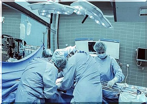
The steps doctors follow to perform a supracondylar amputation are as follows:
- First and foremost, they place the patient on his back.
- Then they mark what is called a fish mouth incision .
- After this, they cut the skin with a cold scalpel.
- Doctors cut the subcutaneous tissue up to the aponeurosis, or mucous membrane that covers the muscles and attaches them to the bone, with an electric scalpel. While the doctor is cutting, there should be enough tissue left for the remaining part of the bone.
- They then identify, isolate and separate the superficial femoral artery and the deep artery in the thigh, as well as the sciatic nerve. It is necessary that they perform a local anesthetic with infiltration.
- They surround the femur and cover the entire circumference.
- After that, the doctors separate the tissues that are attached to the femur. To do this, they must use something called a ” periosteal elevator “.
Final steps
- Physicians should adjust the Percy amputation retractor to amputate the femur. In this way, they can ensure that there is enough soft tissue left to cover the remaining part of the bone.
- After this, the doctors section the leg with a Gigli saw. They do this at an angle of 90 ° between the two ends of the saw. While doing this, they must constantly clean the area with a saline solution.
- Then file the edges of the leg.
- Furthermore, they apply bone wax to the cut part. They press and let the bone wax settle on the cut part. The remains must be thrown away.
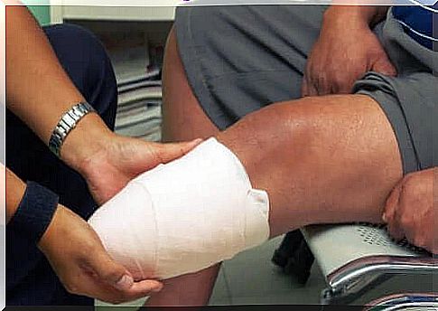
- After this, the doctors close the wound with non-absorbable, sterile surgical sutures, known as Prolene. First, they suture the deeper muscle groups to cover the bone surface. Then they suture the superficial aponeurosis.
- Finally , doctors suture the skin with a silk suture using vertical mattress stitches.